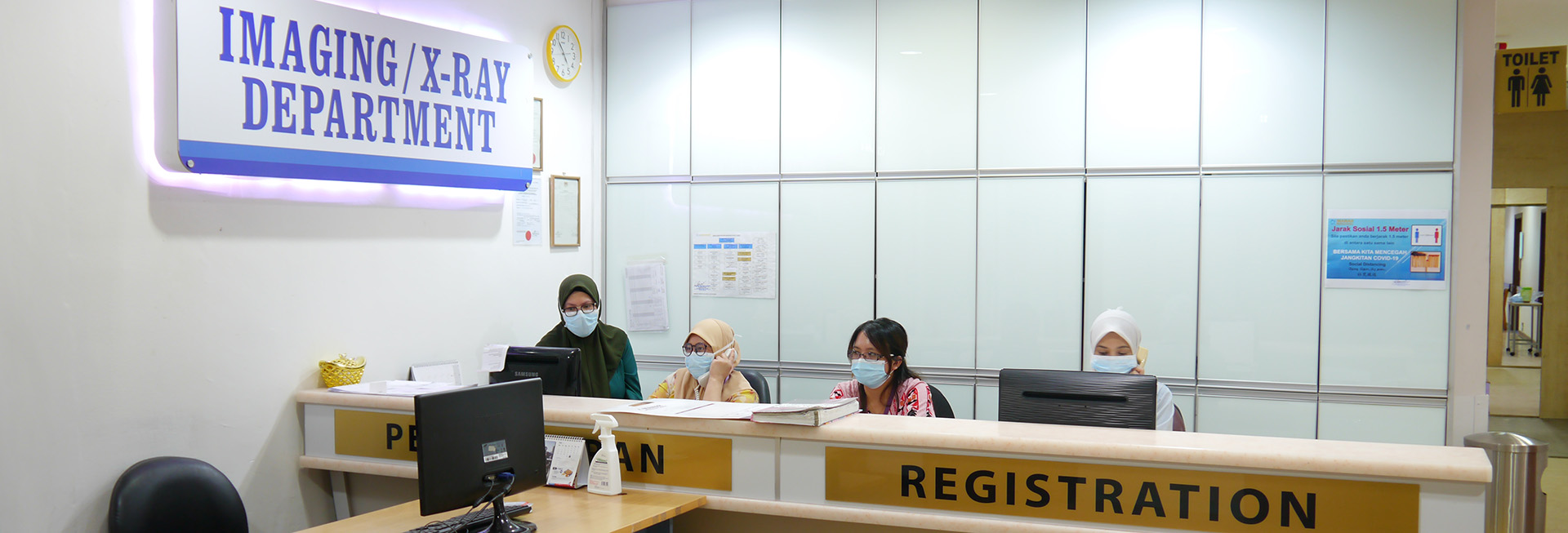

What is MRI?
Magnetic Resonance Imaging (MRI) is a painless and safe diagnostic procedure that uses a powerful magnet and radio waves to produce detailed images of the body's organs and structures, without the use of X-Rays or other radiation.
It helps your doctor to create clear pictures of most parts of the body that give more information than other test such as X-Ray. It is commonly used to get detailed pictures of the brain and spinal cord, to detect abnormalities and tumors.
What is MRI scan and what it used for ?
MRI scan is a medical procedure in which human body organs and structures can be viewed using a large magnet and radio waves.It also is most useful for scanning the brain and spinal cord. It is also used to scan the chest, abdomen, blood vessel and bones.
MRI scan helps to diagnose various medical conditions and diseases. It detects any abnormalities such as tumors, infection, injury, bleeding or diseases of the blood vessels. An MRI scan may also be performed to examine a problem that has been detected in an X-Ray, CT scan or ultrasound scan.
How is the MRI scan examination performed ?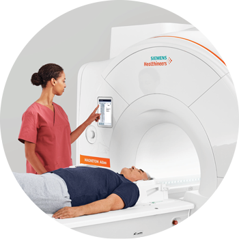
The MRI scan is easy and comfortable. At the start of the scanning process, you will lies on a padded motorized table that slkides in and out of the MRI "tube". Three powerful magnets surround you when positioned inside the MRI tube. Some newer MRI machines are "open" at the sides. Open scanning is of great benefit to claustrophobic you.
The technician operates the MRI machine from a room adjacent to the patient area, which is shared by a large window. The technician is able to see and communicate with the patient during the entire produre. The operation of the equipment from a separate area is necessary to protect the computer from the powerful magnetic forces.
Is MRI scan same with CT scan ?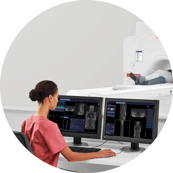
Almost same, but with an MRI scan, it is possible to take pictures form almost every angle, where as a CT scan only shows pictures horizontally. There is no ionizing radiation (X-Rays) involved in producing an MRI scan. MRI scans are generally more detailed. The difference between normal and abnormal tissue is often clearer on the MRI scan than on the CT scan.
Are there any risk in having a MRI scan?
- The MRI examination poses almost no risk to the average patient when appropriate safety guidelines are followed.
- Although the string magnetic field is not harmful in itself, implanted medical devices that contain metal may malfunction or cause problems during an MRI exam.
- There is a very slight risk of an allergic reaction if constrast material is injected. Such reactions usually are mild and easily controlled by medication. If you experience allergic symptoms, a radiologist or other physician will be available for immediate assistance.
- Nephrogenic systemic fibrosis is currently a recognized, but rare, complication of MRI believed to be caused by the injection of high does of gadolinium constrast material in patients with very poor kidney function.
- You may be asked to wear a gown during the exam or you may be allowed to wear your own clothing if it is loose-fitting and has no metal fasteners.
- Guidelines about eating and drinking before an MRI exam vary with the specific exam and also with the facility. For some types of exams, you will be asked to fast for 8-12 hours. Unless you are told. Otherwise, you may follow your regular daily routine and take medications as usual.
- You also required to remove all metal jewelry and electronic devices.
What is Computed Tomography?
A painless test that combines X-Rays and computers processing technology to produce cross sectional images that appear as slices containing detailed images of your internal structures and organs.
It allows your doctor and radiologist to quickly view in extraordinarily fine detail your heart and associated vascular system and other vital organs.
This information helps doctors diagnose a wide variety of conditions earlier and faster than ever before, including your risk for heart and coronary artery disease, as well as other disease or abnormalities
If doctors do see something on your scan, that information can be extremely vital in determining the proper treatment options.
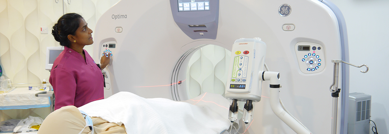
How is the CT Scan examination performed and how long does the CT Scan examination take?
CT exams are quick and comfortable. You will be asked to lie still on a table as it gently moves you through the scanner. Be sure to inform your physician or the technologist if you have any allergies or believe you are pregnant.
The length of your CT exam depends on which particular study, or studies, your doctor has ordered.
Most cardiovascular exams last just a few minutes. You may be asked to arrive at the facility 15 or 30 minutes prior to your scheduled exam time.
Is CT Scan Like X-Ray?
Yes. CT uses X-rays in conjunction with advanced computer technology to generate very accurate and detailed images of your internal organs and structures. Your technologist will step into a control room to conduct the actual exam. You may notice a mechanical noise coming from the scanner. That is just the X-Ray tube being activated and rotating around your body.
Preparation for the CT Scan
Most CT examinations do not usually require any special patient preparations. In some cases the staff may ask you to change into a hospital gown for the exam. You may be asked not to eat or drink anything before your exam. You may also be given a contrast agent (intravenously or to drink) to help highlight a particular organ or body structure.
What is Contrast Agent ?
A contrast agent is a safe liquid substance that makes certain tissues stand out more clearly against their surroundings, enabling the finest details to show up on the X-Ray, improving diagnostic accuracy. You may be given the contrast agent intravenously or orally.
In all cases the contrast agent will leave your body naturally within a few hours. If your exam does require a contrast agent, be sure to tell the technologist if you have any allergies especially to iodine or shellfish.
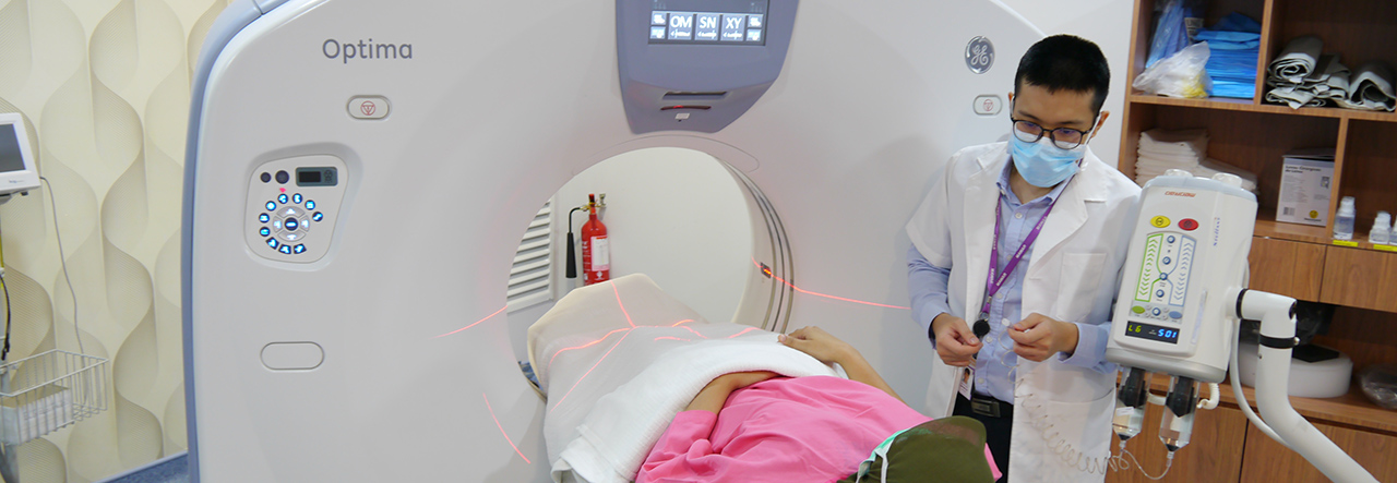
What examination methods are available?
Calcium Scoring is one of several CT scan options that doctors can use to evaluate your cardiovascular health . CT Calcium Scoring is a noninvasive test for quantifying coronary artery calcium content.
The information acquired during your CT exam is processed with a specific cardiac scoring software package that evaluates and quantifies the amount of calcium in your coronary arteries. The calcium content correlates to the degree of blockage in your arteries and consequently provides clinicians with a good indication of what risks you face from heart disease.
Others CT Scan methods available are:
- Coronary CT Angiogram
- Total body vascular scan
- Total body scan
- CT brain
- CT face and neck
- CT chest and lungs
- CT abdomen and pelvis
- CT virtual colonoscopy
Why to choose MRMC's CT Scan?
The C.T. Scanner is capable of performing:
- C.T. Coronary Angiogram
- Coronary Calcium Scoring for Atheroma Assessment

What is Mammogaraphy?
Mammography is a specific type of imaging that uses a low-dose x-ray system to examine breasts. A mammography exam, called a mammogram used to aid in the early detection and diagnosis of breast diseases in women.
An X-Ray (radiograph) is a noninvasive medical test that helps physicians diagnose and treat medical conditions. Imaging with X-Rays involves exposing a part of the body to a small dose of ionizing radiation to produce pictures of the inside of the body. X-Rays are the oldest and most frequently used form of medical imaging.
How is the Mammogram performed and how long does it take?
The mammograms are quick, comfortable and easy. You will be asked to remove all clothing above the waist including jewelry and metal objects from around the neck.
Mammograms are used as a screening tool to detect early breast cancer in women experiencing no symptoms and also to detect and diagnose brease in women experiencing symptoms such as a lump, pain or nipple discharge.
Breast Cancer
- Breast cancer is the most common cancer in women and is a major cause of illness and death.
- If found early, the changes of controlling the disease is high and it may not be necessary to remove the affected breast.
- Signs of breast cancer may include pain, skin thickening, nipple discharge, or a change in breast size or shape.
- At least 40 years old
- Never had a mammogram before or the last mammogram was more than two (2) years old.
- Have any of the following risk factors :-
- Have a mother, sister or daughter with breast cancer
- Menopause and on hormone replacement therapy
- Have had previous history of breast cancer
- Have breast lump(s) or recent changes in the breasts or your doctor has recommended a mammogram for you
- Long-term use of menopausal hormone therapy
- Seek advice early if you notice any change in your breast. Don't wait and worry.
- If you are 40 years and above, require your doctor for a mammogram.
- As your age increases, regular mammograms should become part of your usual health checks.
- Have your doctor examined your breast at least once a year.
- Do monthly breast self examination(BSE).
- Preferably, you should schedule your test one week after your menstrual period.
- Wear a comfortable 2-piece outfit.
- Do not use deodorant, perfume, powder or ointment on the underarms or breast. They may produce artifacts in the radiographs
- On the day of the exam, wear a skirt or pants, rather that a dress, since you will need to remove your top for the test.
- Avoid scheduling your mammogram at a time when your breasts are swollen or tender, such as right before your period.
What is a Densitometry?
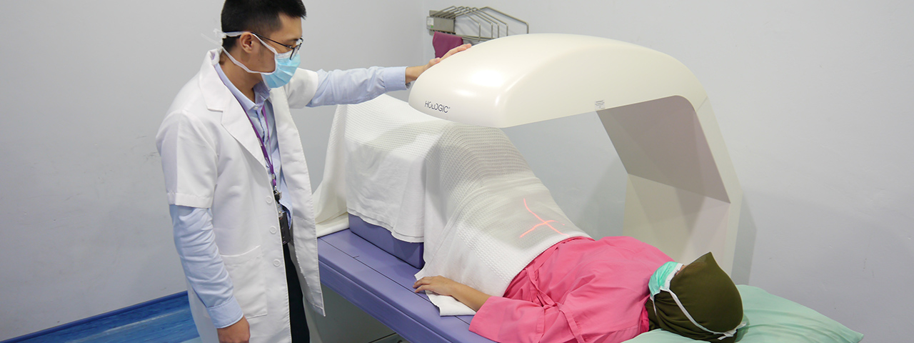
Densitometry is a special type of X-ray test used to measure the calcium content of the bone. The examination is also called a Dual Energy X-ray Absorptiometry (DEXA) scan or QDR scan. The DEXA scan is the established standard for measuring Bone Mineral Density (BMD).
This is a simple, quick and non-invasive medical test, which involves exposing particular parts of the body to very small amounts of ionisation radiation. This is then used to produce images of the insides of the body. DEXA scans are used to measure and therefore diagnose low bone density and also to monitor patients where a diagnosis of low bone density has been made. DEXA scans are usually performed on the lower spine and the hips.
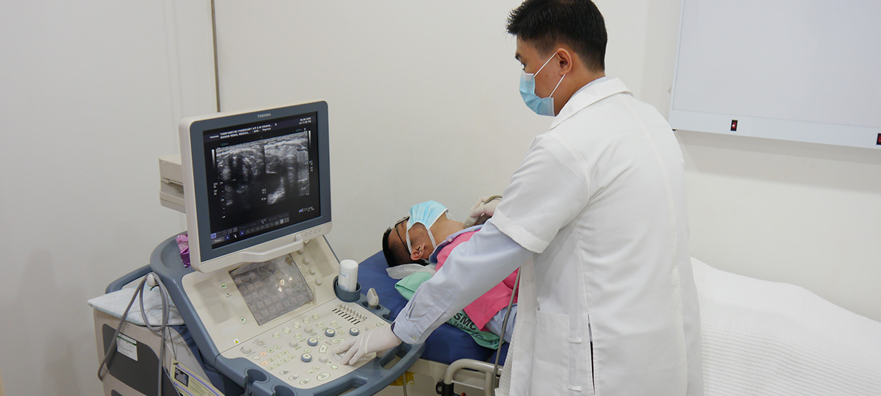
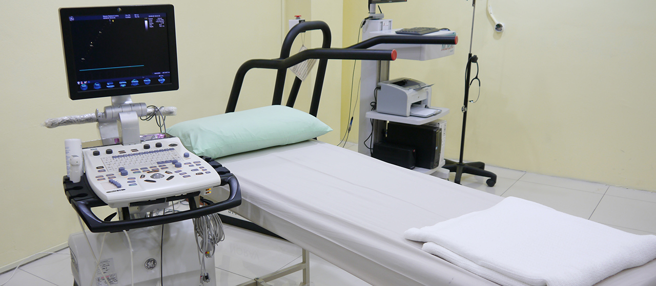
An ultrasound is an imaging test that uses sound waves to make pictures of organs, tissues, and other structures inside your body. It allows your health care provider to see into your body without surgery. Ultrasound is also called ultrasonography or sonography. Ultrasound images may be called sonograms.
-
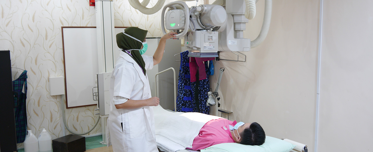
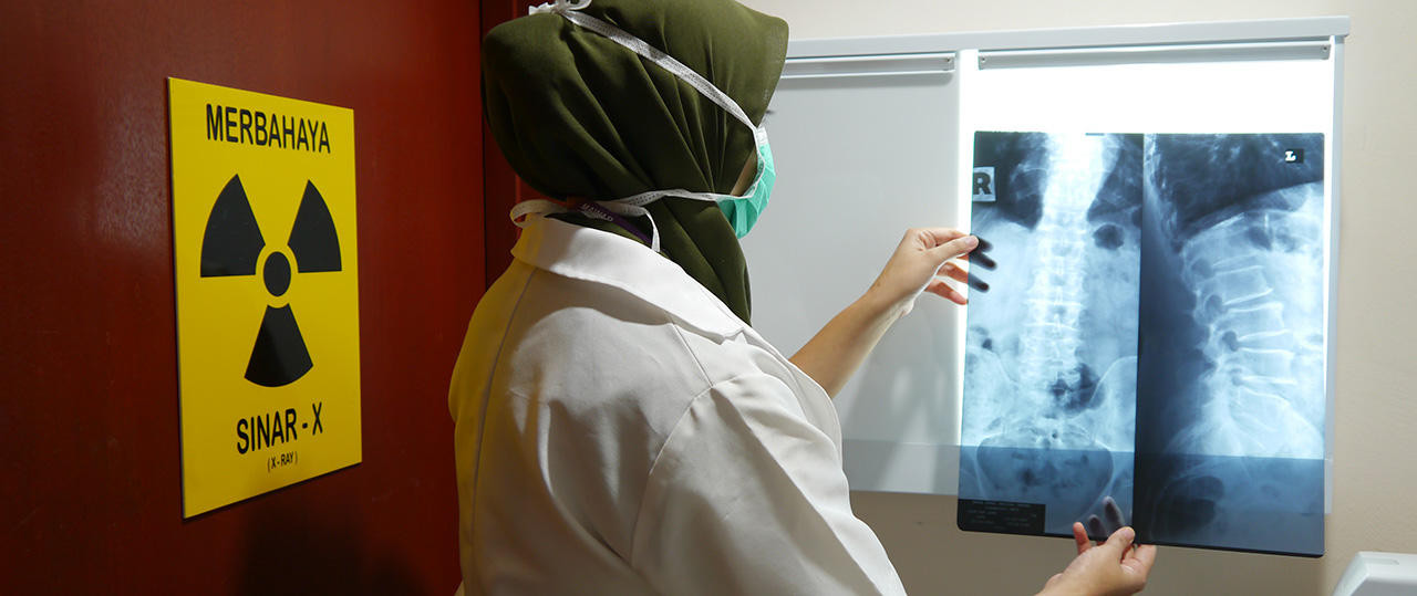
X-Rays are a form of radiation, like light or radio waves, that can be focused into a beam, much like a flashlight beam. Unlike a beam of light, however, X-Rays can pass through most objects, including the human body.
- When X-Rays strike a piece of photographic film, they can produce a picture. Dense tissues in the body, such as bones, block (absorb) many of the X-Rays and appear white on an X-Ray picture. Less dence tissues, such as muscles and organs, block fewer of the X-Rays (more of the X-Rays pass through) and appear in shades of gray. X-Rays that pass only through air appear black on an X-Ray picture.
What is Electrocardiogram (ECG)?
- The electrocardiogram (ECG) is a noninvasive test that is used to reflect underlying heart conditions by measuring the electrical activity of the heart.
- By positioning leads (electrical sensing devices) on the body in standardized locations, information about many heart conditions can be learned by l;ooking for characteristic patterns.
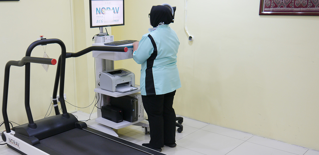
Exercise stress testing is tremendously informative in screening for coronary artery disease as well as periodically following known heart disease.

